


Author: Diva John
Mentor: Dr. Edina Poletto
Lotus Valley International School Gurugram
Abstract
It has been known that pregnancy is a profound biological transformation where the maternal body can reshape its cardiovascular, physiological, and immune system to sustain the developing fetus.
Such intense anatomical restructuring, especially neurologically, can leave women highly vulnerable to medical complications both during gestation and in the immediate postpartum state.
Tremendous surges of estrogen, progesterone, and placental cortisol actively contort and restructure maternal neural networks to prepare the brain for the postnatal period.
Postpartum psychosis is a severe mental health condition which affects people after they give birth. This condition is rare and dangerous. People with postpartum psychosis have a much higher risk of harming themselves, dying by suicide or harming their children.
However, the hypothesis that a sudden postpartum crash in these chemical messengers can cause maladaptive neuroplasticity, severely disrupting circuits to trigger postpartum depression or postpartum psychosis is worth investigating to control any such cases. The hormonal shifts in the mother’s brain, as well as the extent of altered neuroplasticity, would have to be monitored to ensure the right amount of adaptive restructuring after pregnancy.
Keywords: hormones, brain alteration, structural changes, functional consequences, maternal maladaptation, Post-partum psychosis
Introduction
During the eighteenth century, first found records of Friedrich Hoffman, citing the appearance of a condition same as postpartum psychosis was observed in his book
Medicinae Rationalis Systematicae (Systematic Rational Medicine). Almost 400 non-organic episodes in the literature started more than 3 weeks after the birth, some of them being recurrent.
Hoffmann described post-partum psychosis symptoms in a young woman as following (1721): [Brockington I, 2017]
“Four weeks after childbirth a 20-year old, rather melancholy and anxious by temperament, had a severe fright – she ‘saw’ the ghost of her long-dead mother. Three days later she became confused and started to rave. She was restless, talked day and night (mainly in rhyme), and ate and drank little, but had no fever. The infant was given to the wet-nurse, leeches were applied to her feet, and medicines given to bring on the menses; but her raving increased more and more, and she had to be restrained. It was 2 months before she calmed down. She immediately became pregnant again and remained well.”
The first case series were seen in the 19th century out of which Esquirol’s statements were the most detailed. Postpartum psychosis symptoms were observed in 92 women of which around 53% of the women predominantly contained manic symptoms, 38% showed depressive symptoms, and 9% of them displayed nonaffective psychosis. [Bergink V et al., 2016]
The connections between parturition and post-partum psychosis has been overlooked historically, despite them having hypothesized a definite pathophysiology path. Although there is a connection observed, the cellular mechanisms and the fundamental basics of the molecular changes that occur during post-partum psychosis still remain quite a mystery.
During pregnancy, the maternal placenta and the fetus’s endocrine system work together to manage the endocrine changes that are required to maintain and promote pregnancy and also to prepare women for childbirth, breastfeeding, and other important processes post-partum. [Stern C et al., 2021] Majority of the endocrine hormones are sex hormones, example- estrogens and progesterone, that steadily increase throughout pregnancy having peaks during certain months and drastically fall at parturition. Progesterone is largely produced by the corpus luteum until about 10 weeks of gestation. The powerful influence of estrogens and progesterone (along with other hormones) on the neuroanatomical state of the brain is demonstrated during puberty, menopause, the menstrual cycle and pregnancy. Therefore, consequential changes in the brain’s physical state are also inevitable during pregnancy and after giving birth. However, research in this field is relatively scarce. [Kumar P et al., 2012]
Some symptoms of postpartum psychosis are insomnia, irritability and mood shifts, with an increase in mania, depression, or even a mixed state. Rapid mood fluctuations are a characteristic visual of the disease, however women suffering from postpartum psychosis have symptoms that are often atypical in patients with bipolar disorder. [Osborne LM et al., 2018] Disorganized, unusual behavior and obsessive thoughts with relation to the newborn occur frequently. Various cognitive symptoms such as disorientation, confusion, derealization, and depersonalization may also come along with post-partum psychosis. Delusions of altruistic homicide (seen with maternal suicide at times) to “save them both” from a fate apparently worse than death may occur and are an important exploration within the clinical examination. PPP is attached with an increased risk of both suicide and infanticide. [Bergink V et al., 2016]
Although many longitudinal neuroimaging studies have established that pregnancy is accompanied by extensive neuroanatomical remodeling, the functional importance and adaptive extent of these changes remain incompletely understood.
The question comes whether the preservation of adaptive maternal neuroplasticity through modulation of perinatal stress and early identification of aberrant neural trajectories will reduce vulnerability to maladaptive maternal behavior and peripartum mental disorders. [ L Pritschet et al., 2024]
Neuroanatomical Remodeling During Pregnancy
Grey matter volume and cortical thickness change
Reductions have been observed in the volume of grey matter in the brain (GMV) post childbirth, especially in the regions of the brain central to theory-of-mind- processing. Such changes could be visible even after 6 years postpartum and can be traced decades later.[Losse EM et al., 2026] These findings depict that pregnancy is an increasingly vigorous stage for neural restructuring, and currently neuroscientists lack a detailed map of how exactly the human brain shifts its state throughout the period of gestation. [ L Pritschet et al., 2024]
Both the cortical thickness and grey matter decrease has been observed throughout pregnancy, and after birth has partially jumped back. The cerebral cortex and other large-scale brain networks experienced grey matter volume increase across their regions. Even in certain parts of the brain deep within, there was a decrease in grey matter volume. All these changes were substantially larger in pregnant women, as compared to the women who were not pregnant over a similar period of observation.
White matter is another integral tissue in the brain consisting mainly of nerve fiber tracts which carry signals between the regions of grey matter in the brain. The fine-scale health and packing density of white matter here increased the entirety of the first two trimesters of pregnancy. This increase returned to its baseline level after birth of the child. Volumes of cerebrospinal fluid in the lateral ventricles (C-shaped cavities) had also increased during the second and third trimesters of pregnancy, which then dropped sharply after birth. These brain changes observed were associated with shifts in steroid hormone levels. This supports the idea that pregnancy is a period of intensive neuroplasticity, a capacity of various networks of neurons to adapt and reorganize fast. [ L Pritschet et al., 2024]
Subcortical changes
In tandem with reductions in broader cortical regions in GMV, various subcortical regions showed significant reduction in volume across gestation(fig 1, left). This included the bilateral ventral diencephalon ( right hemisphere values depicted in fig.1, right; compromising hypothalamus, substantia nigra, mammillary body, lateral geniculate nucleus and red nucleus among others), caudate, hippocampus and thalamus, along with left putamen and brain stem. [Pritschet L et al., 2024]
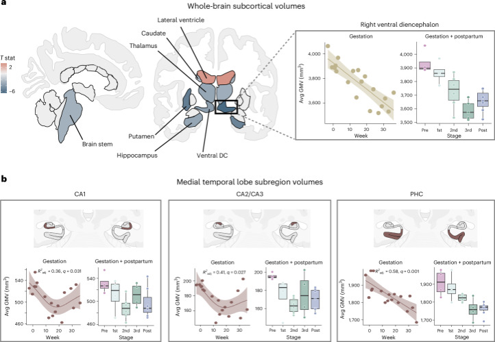
Fig. 1. Subcortical GMV changed throughout gestation, adapted from ( Pritschet L, 2024)
Mechanisms of Maternal Neuroplasticity
Hormone action
Pregnancy activates intense physiological and neurobiological shifts in maternal body and brain, including a sharp rise in movement of sex steroid hormones, especially estrogen and progesterone. [Losse EM et al., 2026]
Due to the lipid-like nature of steroid hormones, certain hormones like progesterone and estradiol passively cross the blood brain barrier which lets them bind to the estrogen and progesterone receptors present in the brain. Such steroids act on a network of hormone-sensitive regions of the brain which are identified as maternal caregiving networks. [Catenaccio E et al., 2016] These shifts play an important role in making the maternal brain fit for childcare, regulating neurotransmission and neuroplasticity by interactions with the key receptor systems, including primary inhibitory and excitatory neurotransmitters.
GABA Synthesis and working
Gamma-aminobutyric acid (GABA) and glutamate (N-methyl-D-aspartate receptor; NMDA), are principal to neuroplasticity processes. Estrogen catalyses glutamatergic neurotransmission by increasing the NMDA receptor subunit expression, the number of binding sites, and the neuronal sensitivity to synaptic input. [Sears SM et al., 2021]
Allopregnanolone is a potent positive modulator of GABAA receptors and it helps with the excitatory/inhibitory balance (E-I Balance) which is required for maintaining neural stability. The simultaneous release of GABAA and glutamate is essential for brain functioning, particularly in regions like the prefrontal cortex, amygdala and the hippocampus. It plays a vital role here in cognitive and emotional regulation. In certain animal models, GABAA and glutamate have been observed to get synchronously upregulated during postpartum, depicting a coordinated neurochemical adaptation for motherhood. In mice, dysregulation or imbalance in E-I after a simulated pregnancy was associated with depression-like behaviour, and restoration of the E-I balance mitigated the condition. [Losse EM et al., 2026]
Oxytocin
Beside these neurotransmitter systems, oxytocin comes out as another important modulator of maternal brain working. It is a neuropeptide which enhances the synaptic excitability and promotes neuroplasticity, fortifying maternal behavior. [Froemke RC et al., 2021]Oxytocin levels increase progressively with mother-infant interactions, further improving their attachment.[Scatliffe N et al., 2019] It also interacts closely with the GABAergic system, implying its broader role in regulating mood regulations and showing resilience to stress in the postpartum period. [Walter MH et al., 2021]
HPA-Axis
The hypothalamic-pituitary-adrenal (HPA) axis adapts during pregnancy and postpartum to remake the maternal brain. Glucocorticoid and corticotropin-releasing hormone (CRH) signaling undergo alterations due to HPA-axis. These changes facilitate structural and functional neuroplasticity in brain regions which control anxiety, reward, and maternal caregiving. Altered fetal HPA axis ultimately results in dysregulation of the brain stress-response system long after birth and possibly lifelong. [O’Keane V et al., 2011]
Functional Significance of Maternal Brain Remodeling
Mother-infant interactions based on neural changes
The structural and functional plasticity in the human maternal brain is associated with individual differences in maternal thoughts and behaviors. With reference to structural plasticity, it is seen that the greater the growth in the midbrain region, the higher the positive emotions a mother reports about her baby, which could indicate positive adaptation to parenthood in the third and fourth months postpartum. [Kim P , 2016]
At 4 to 6 months postpartum, neural activation patterns associated with sensitive and synchronous maternal behaviors (for example: increased coordination of gaze, touch, and vocalization with infants) were examined. In response to a mother’s own baby video stimuli as compared to control baby video stimuli, synchronous mothers had greater activation in the nucleus accumbens, which is a key reward/motivation region. This functionally correlated with activity in the inferior frontal gyrus as well as medial frontal gyrus. Thus, reward-related neural responses to one’s own infant were associated with magnified neural connectivity for social information processing, which may further support mother-infant interactions.
Maternal neural responses to infants at the time of early postpartum can play a crucial role to predict positive infant socioemotional results. Among a few new mothers, high levels of anxious thoughts in the mother about their own infants and their competency as a parent at the first month postpartum showed lower socioemotional competencies among their infants at 18-24 months of age. High anxiety about parenting and infants was further associated with decrease in activation in the substantia nigra,, in response to their own infant cry compared to other infant cry sounds at the first month postpartum, suggesting reduced neural sensitivity to own infants. [Kim P, 2016]
These understandings also suggest that abnormality in the neural plasticity of a maternal brain could increase vulnerability for a difficult transition to parenting among some mothers. Mothers may be vulnerable to severe difficulties in mood regulation and stress. New mothers are exposed to increased levels of risk for mood disorders like postpartum depression and anxiety disorders. [Garapati J et al., 2023]
Together, the findings from these studies show a structural increase in many maternal brain regions that are involved in parenting, from immediately after childbirth to 3–4 months postpartum.
Dysregulated Neuroplasticity and Maladaptive Outcomes
The decrease observed in GMV, brain volume, neurogenesis and glial density can actually be part of the normal optimization of maternal behaviour, rather than deterioration (JL. Pawluski 2022). So the matter to understand is how the same broad phenomenon of neuroplastic remodeling is adaptive in one context but potentially maladaptive in another.
Some possible agents that could deviate the normal optimal working of the maternal brain due to its changes are harmful stressors, hormonal dysregulations and the most underscored one yet, E/I imbalance in the brain.
Stress
Stress has been introduced by Hans Selye as the GAS, which is a process in which the body confronts noxious agents. The mechanisms in which external stressors alter brain function have been studied extensively over the past 60 years. [Rochette L et al., 2023] Corticotropin-releasing hormone (CRH), the stress hormone, has become the focus of the study. In both animals and humans, CRH stimulates norepinephrine (NE) synthesis and release. CRH release activates the hypothalamic-pituitary-adrenal (HPA) axis, which acts as one of the main mechanisms involved in stress. The CRH induced synthesis of ACTH (adrenocorticotropic hormone) in turn alters the function of the neural network by changing the building blocks in the networks and by restructuring the integrative properties, finally causing the behavioral or emotional changes. [Contoreggi C et al., 2015] This neural plasticity underlying stress, undoubtedly impacts the brain function and can cause functional alterations in mental disorders. Thus, stress-induced neuroplasticity plays a pivotal role in almost all of the mental disorders, and has become a synonym for displaying certain negative emotions, such as depression and anxiety.
Hormone dysregulation
The hormonal changes attached with pregnancy can attain levels which could mimic diseases like cushing disease, diabetes mellitus, hyperthyroidism and more, these themselves being associated with psychiatric disturbances. Such hormonal shifts can also alter the circadian rhythm of the body causing negative impact on health and leading to insomnia. Further, Insomnia is known to be associated with depression and suicidal ideation. [Trifu S et al., 2019]
The sudden fall in progesterone post-partum can have a role in postpartum depression. An explanation hypothesised is that progesterone and its cerebral metabolite called allopregnanolone decrease irritability. Ovaries don’t secrete progesterone again until the first menstrual cycle occurs, which can cause a temporary imbalance. The brain and body adapts to low levels of estradiol which promote lactation. Estradiol works to facilitate serotonergic transmission by catalysing synthesis of serotonin and/or reducing reuptake of serotonin, thus reducing depressive symptoms. The reduced estradiol levels in the maternal body have the opposite effect, depriving it of its natural protection against depression. [Hernández-Hernández OT et al., 2019]
Testosterone is known to have a relation to greater mood disturbances after birth.
Plasma corticosteroids reach a high during labor and reduce gradually within 4 hours postpartum, creating an emotionally vulnerable period for the mother.In the presence of low estradiol, potentially lower dopamine and variations in thyroid hormones, it is not uncommon that such imbalances can cause an important psychological impact. [Trifu S et al., 2019]
E/I Imbalance
Persistent E/I imbalance could be one mechanism distinguishing normal postpartum neuroplasticity from maladaptive neuroplasticity. The E/I balance is impactful enough to cause the shift from normal neural stability to depression like behaviour. [ Yin, Y. Y., et al., 2021] If in certain cases, this balance is not restored due to insufficient GABA receptors or excess glutamate, deregulation of the E-I balance could cause a number of nervous system disorders, such as epilepsy, mental retardation and autism. Prolonged imbalance could alter the extent of neuroplasticity of the maternal brain, and cause episodes similar to those seen in postpartum-psychosis.
Preventive and Therapeutic Implications
If stress-related dysregulation contributes to abnormal neuroplasticity, interventions that improve regulation of the stress system might indirectly protect adaptive brain remodeling.
As normal plasticity is adaptive and dysregulated plasticity becomes potentially harmful, then a possible control for preventing maladaptive outcomes is by controlling either the extent of plasticity or the imbalance that can occur in the maternal brain before parturition occurs. During normal pregnancy, the maternal stress response system itself changes. Severe stress can disrupt this adaptation. [Coussons-Read ME, 2013]
Other possibilities that can be used to scope out treatments include finding the abnormal neural trajectories. Longitudinal MRI’s of the maternal brain that get monitored during and after parturition and landscaping the paths and changes of the brain that shift from the normal trajectories are ways post-partum disorders can be prevented and controlled. [Kim P et al., 2010] Mothers who are giving birth and have any previous exposure to certain substances, stressors, or early psychological trauma must be checked regularly to ensure that slight changes don’t alter the makeup of the brain, potentially causing post-partum psychosis or post-partum depression. If any such patients are observed then reducing the possibilities of any exposure to such stressors must be eliminated.
E/I Imbalance is an underlying problem, which if not restored, can create maladaptive pathways to brain abnormalities which cause manic and mood shift behaviours. This is still largely a future-research proposition, not an established clinical screening method. Postpartum psychosis is a medical emergency which requires immediate psychiatric hospitalization. The best current approach involves fast-acting antipsychotics and benzodiazepines for acute symptom control, the mood stabilizer lithium as the gold standard for long-term recovery and prevention, and adopting electroconvulsive therapy (ECT) for severe or extremely resistant cases. [Jairaj C et al., 2023] However, prevention is much better than draining the body out with cures, so before ppp occurs, for any female with the chance of having post-partum disorders or facing immense stress, close monitoring is the best safety measure.
Abbreviations
- GMV- Grey matter volume
- PPP- Post-partum psychosis
- GABA- Gamma amino butyric acid
- NMDA- N-methyl-D-Aspartate receptor
- E/I- Excitatory/Inhibitory
- HPA- Hypothalamic-pituitary-axis
- CRH- Corticotropin-releasing hormone
- GAS- General adaptation syndrome
- NE- Norepinephrine
- ACTH- Adrenocorticotropic hormone
- MRI- Magnetic resonance imaging
- ECT- Electroconvulsive therapy
References and Citations
Altemus, M., Redwine, L. S., Leong, Y.-M., Frye, C. A., Porges, S. W., & Carter, C. S. (2001). Responses to laboratory psychosocial stress in postpartum women. Psychosomatic Medicine, 63, 814–821. https://doi.org/10.1097/00006842-200109000-00015
Atzil, S., Hendler, T., & Feldman, R. (2011). Specifying the neurobiological basis of human attachment: Brain, hormones, and behavior in synchronous and intrusive mothers. Neuropsychopharmacology, 36(13), 2603–2615. https://doi.org/10.1038/npp.2011.172
Been, L. E., Sheppard, P. A. S., Galea, L. A. M., & Glasper, E. R. (2022). Hormones and neuroplasticity: A lifetime of adaptive responses. Neuroscience & Biobehavioral Reviews, 132, 679–690. https://doi.org/10.1016/j.neubiorev.2021.11.029
Bergink, V., Rasgon, N., & Wisner, K. L. (2016). Postpartum psychosis: Madness, mania, and melancholia in motherhood. American Journal of Psychiatry, 173(12), 1179–1188. https://doi.org/10.1176/appi.ajp.2016.16040454
Brockington, I. (2017). Late onset postpartum psychoses. Archives of Women’s Mental Health, 20(1), 87–92. https://doi.org/10.1007/s00737-016-0680-y
Catenaccio, E., Mu, W., & Lipton, M. L. (2016). Estrogen- and progesterone-mediated structural neuroplasticity in women: Evidence from neuroimaging. Brain Structure and Function, 221(8), 3845–3867. https://doi.org/10.1007/s00429-016-1197-x
Contoreggi, C. (2015). Corticotropin releasing hormones and imaging, rethinking the stress axis. Nuclear Medicine and Biology, 42(4), 323–339. https://doi.org/10.1016/j.nucmedbio.2014.11.008
Coussons-Read, M. E. (2013). Effects of prenatal stress on pregnancy and human development: Mechanisms and pathways. Obstetric Medicine, 6(2), 52–57. https://doi.org/10.1177/1753495X12473751
Del Río, J. P., Alliende, M. I., Molina, N., Serrano, F. G., Molina, S., & Vigil, P. (2018). Steroid hormones and their action in women’s brains: The importance of hormonal balance. Frontiers in Public Health, 6, 141. https://doi.org/10.3389/fpubh.2018.00141
Eichler, S. A., & Meier, J. C. (2008). E-I balance and human diseases—from molecules to networking. Frontiers in Molecular Neuroscience, 1, 2. https://doi.org/10.3389/neuro.02.002.2008
Elyada, Y. M., & Mizrahi, A. (2015). Becoming a mother—Circuit plasticity underlying maternal behavior. Current Opinion in Neurobiology, 35, 49–56. https://doi.org/10.1016/j.conb.2015.06.007
Florio, A. D., Munk-Olsen, T., & Bergink, V. (2016). The birth of a psychiatric orphan disorder: Postpartum psychosis. The Lancet Psychiatry, 3(6), 502. https://doi.org/10.1016/S2215-0366(16)30037-2
Froemke, R. C., & Young, L. J. (2021). Oxytocin, neural plasticity, and social behavior. Annual Review of Neuroscience, 44, 359–381. https://doi.org/10.1146/annurev-neuro-102320-102847
Garapati, J., Jajoo, S., Aradhya, D., Reddy, L. S., Dahiphale, S. M., & Patel, D. J. (2023). Postpartum mood disorders: Insights into diagnosis, prevention, and treatment. Cureus, 15(7), e42107. https://doi.org/10.7759/cureus.42107
Hernández-Hernández, O. T., Martínez-Mota, L., Herrera-Pérez, J. J., & Jiménez-Rubio, G. (2019). Role of estradiol in the expression of genes involved in serotonin neurotransmission: Implications for female depression. Current Neuropharmacology, 17(5), 459–471. https://doi.org/10.2174/1570159X16666180628165107
Jairaj, C., Seneviratne, G., Bergink, V., Sommer, I. E., & Dazzan, P. (2023). Postpartum psychosis: A proposed treatment algorithm. Journal of Psychopharmacology, 37(10), 960–970. https://doi.org/10.1177/02698811231181573
Kim, P., Leckman, J. F., Mayes, L. C., Feldman, R., Wang, X., & Swain, J. E. (2010). The plasticity of human maternal brain: Longitudinal changes in brain anatomy during the early postpartum period. Behavioral Neuroscience, 124(5), 695–700. https://doi.org/10.1037/a0020884
Kim, P. (2016). Human maternal brain plasticity: Adaptation to parenting. New Directions for Child and Adolescent Development, 2016(153), 47–58. https://doi.org/10.1002/cad.20168
Kim, P. (2021). How stress can influence brain adaptations to motherhood. Frontiers in Neuroendocrinology, 60, 100875. https://doi.org/10.1016/j.yfrne.2020.100875
Kumar, P., & Magon, N. (2012). Hormones in pregnancy. Nigerian Medical Journal, 53(4), 179–183. https://doi.org/10.4103/0300-1652.107549
Losse, E. M., Daneshnia, N., Dukart, J., Nehls, S., & Chechko, N. (2026). Maternal brain alterations based on neurotransmitter and hormone receptor distributions over six months postpartum. Translational Psychiatry, 16(1), 280. https://doi.org/10.1038/s41398-026-04104-4
Lucia, M., Viviana, M., Alba, C., Giulia, D., Carlo, D. R., Porpora, M. G., Luca, T., Federica, V. M., Immacolata, V. A., & Grazia, P. M. (2023). Neurological complications in pregnancy and the puerperium: Methodology for a clinical diagnosis. Journal of Clinical Medicine, 12(8), 2994. https://doi.org/10.3390/jcm12082994
Luders, E., Kurth, F., & Sundström Poromaa, I. (2022). The neuroanatomy of pregnancy and postpartum. NeuroImage, 263, 119646. https://doi.org/10.1016/j.neuroimage.2022.119646
Martínez-García, M., et al. (2021). Do pregnancy-induced brain changes reverse? The brain of a mother six years after parturition. Brain Sciences, 11, 168.
O’Keane, V., Lightman, S., Patrick, K., Marsh, M., Papadopoulos, A. S., Pawlby, S., Seneviratne, G., Taylor, A., & Moore, R. (2011). Changes in the maternal hypothalamic-pituitary-adrenal axis during the early puerperium may be related to the postpartum “blues.” Journal of Neuroendocrinology, 23(11), 1149–1155. https://doi.org/10.1111/j.1365-2826.2011.02139.x
Osborne, L. M. (2018). Recognizing and managing postpartum psychosis: A clinical guide for obstetric providers. Obstetrics and Gynecology Clinics of North America, 45(3), 455–468. https://doi.org/10.1016/j.ogc.2018.04.005
Pawluski, J. L., Hoekzema, E., Leuner, B., & Lonstein, J. S. (2022). Less can be more: Fine tuning the maternal brain. Neuroscience & Biobehavioral Reviews, 133, 104475. https://doi.org/10.1016/j.neubiorev.2021.11.045
Rochette, L., Dogon, G., & Vergely, C. (2023). Stress: Eight decades after its definition by Hans Selye: “Stress is the spice of life.” Brain Sciences, 13(2), 310. https://doi.org/10.3390/brainsci13020310
Ruffaner-Hanson, C., Noor, S., Sun, M. S., Solomon, E., Marquez, L. E., Rodriguez, D. E., Allan, A. M., Caldwell, K. K., Bakhireva, L. N., & Milligan, E. D. (2022). The maternal-placental-fetal interface: Adaptations of the HPA axis and immune mediators following maternal stress and prenatal alcohol exposure. Experimental Neurology, 355, 114121. https://doi.org/10.1016/j.expneurol.2022.114121
Scatliffe, N., Casavant, S., Vittner, D., & Cong, X. (2019). Oxytocin and early parent-infant interactions: A systematic review. International Journal of Nursing Sciences, 6(4), 445–453. https://doi.org/10.1016/j.ijnss.2019.09.009
Sears, S. M., & Hewett, S. J. (2021). Influence of glutamate and GABA transport on brain excitatory/inhibitory balance. Experimental Biology and Medicine, 246(9), 1069–1083. https://doi.org/10.1177/1535370221989263
Sit, D., Rothschild, A. J., & Wisner, K. L. (2006). A review of postpartum psychosis. Journal of Women’s Health, 15(4), 352–368. https://doi.org/10.1089/jwh.2006.15.352
Spalek, K., et al. (2024). Pregnancy renders anatomical changes in hypothalamic substructures of the human brain that relate to aspects of maternal behavior. Psychoneuroendocrinology, 164, 107021.
Stern, C., Schwarz, S., Moser, G., Cvitic, S., Jantscher-Krenn, E., Gauster, M., & Hiden, U. (2021). Placental endocrine activity: Adaptation and disruption of maternal glucose metabolism in pregnancy and the influence of fetal sex. International Journal of Molecular Sciences, 22(23), 12722. https://doi.org/10.3390/ijms222312722
Trede, K., Baldessarini, R. J., Viguera, A. C., & Bottero, A. (2009). Treatise on insanity in pregnant, postpartum, and lactating women (1858) by Louis-Victor Marcé: A commentary. Harvard Review of Psychiatry, 17(2), 157–165. https://doi.org/10.1080/10673220902891802
Trifu, S., Vladuti, A., & Popescu, A. (2019). The neuroendocrinological aspects of pregnancy and postpartum depression. Acta Endocrinologica (Bucharest), 15(3), 410–415. https://doi.org/10.4183/aeb.2019.410
Walter, M. H., Abele, H., & Plappert, C. F. (2021). The role of oxytocin and the effect of stress during childbirth: Neurobiological basics and implications for mother and child. Frontiers in Endocrinology, 12, 742236. https://doi.org/10.3389/fendo.2021.742236
Wang, F., Pan, F., Shapiro, L. A., & Huang, J. H. (2017). Stress induced neuroplasticity and mental disorders. Neural Plasticity, 2017, 9634501. https://doi.org/10.1155/2017/9634501
Xu, K., Song, D., Li, H., Lu, Y., & Zhu, Z. (2025). Advance in the mechanisms underlying prenatal stress-induced depressive-like behavior in offspring. Brain, Behavior, & Immunity—Integrative, 9, 100108. https://doi.org/10.1016/j.bbii.2025.100108
Yin, Y. Y., Wang, Y. H., Liu, W. G., Yao, J. Q., Yuan, J., Li, Z. H., Ran, Y. H., Zhang, L. M., & Li, Y. F. (2021). The role of the excitation–inhibition functional balance in the mPFC in the onset of antidepressants. Neuropharmacology, 191, 108573. https://doi.org/10.1016/j.neuropharm.2021.108573
Zoubovsky, S. P., Hoseus, S., Tumukuntala, S., Schulkin, J. O., Williams, M. T., Vorhees, C. V., & Muglia, L. J. (2020). Chronic psychosocial stress during pregnancy affects maternal behavior and neuroendocrine function and modulates hypothalamic CRH and nuclear steroid receptor expression. Translational Psychiatry, 10(1), 6. https://doi.org/10.1038/s41398-020-0704-2
About the author
Diva John
Diva is an incoming medical student from India with a strong interest in neuroscience, psychology, and the biological mechanisms underlying human health and behaviour. Her research interests particularly lie in neuroplasticity and emerging areas of neuroscience that remain relatively underexplored.
She has also previously gained an understanding of clinical exposure through a psychology internship at Fortis Hospital. Through her research, Diva hopes to continue exploring questions at the intersection of neuroscience, psychology, and medicine.